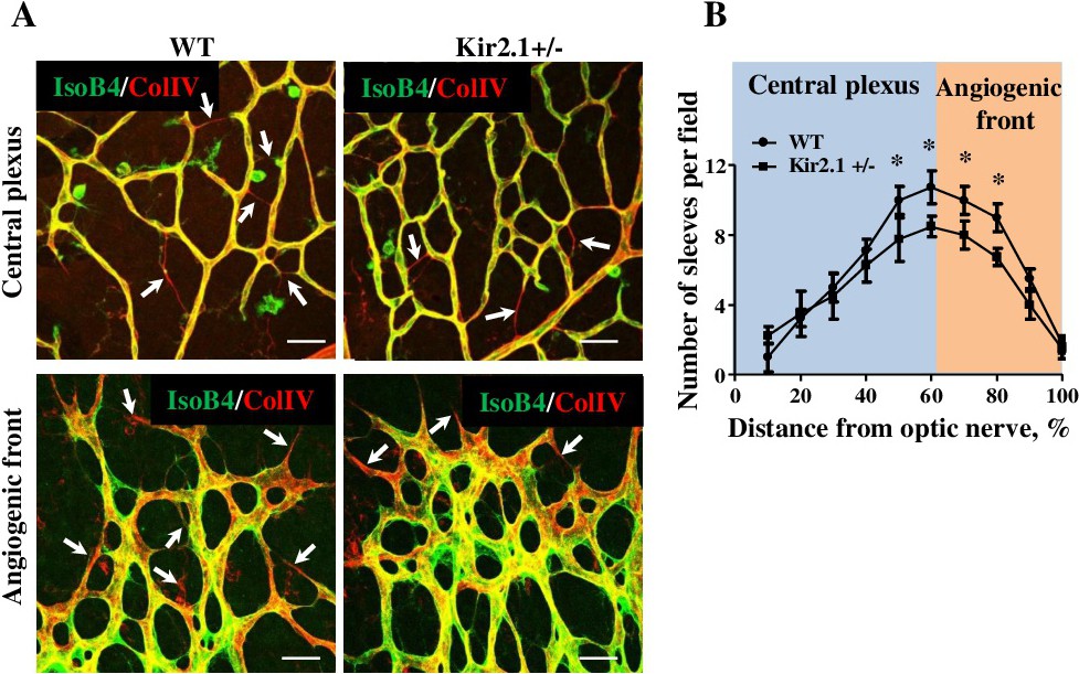
Fig. 4. Kir2.1 deficiency leads to delay in vascular remodeling. (A) Images of retinas of WT and Kir2.1+/- mice stained for IsoB4 (green) and collagen IV (red). Collagen positive and isolectin B4 negative indicate empty sleeves that remain after remodeling. (B) Quantification of empty sleeves across the retina in wild type and Kir2.1+/- retina. Data are means ± SD of at least six mice per group. *P˂0.05. Scale bar panel B: 50 µm.